July 7, 2026
Keratoconus: When the Cornea Changes Shape
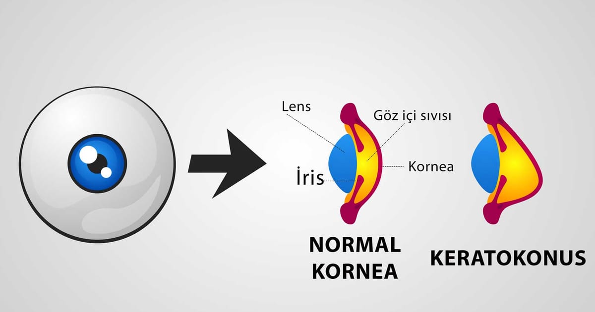
The cornea — the clear front window of the eye — is normally shaped like a smooth dome. In keratoconus, it gradually thins and bulges outward into an irregular cone. That change scatters light and distorts vision in a way ordinary glasses struggle to correct. Keratoconus usually begins in the teens or twenties and can slowly progress for years, which is why recognising it early matters so much.
What causes keratoconus?
The exact cause is not fully understood, but it involves a weakening of the cornea's internal structure. Several factors are linked to it:
- Genetics — it can run in families
- Vigorous, habitual eye rubbing, which is strongly associated with progression
- Allergic eye disease and constant itching
- Certain conditions such as Down syndrome and connective-tissue disorders
Because eye rubbing is a modifiable factor, protecting the eyes from allergy and breaking the rubbing habit are among the simplest ways to help slow the disease.
Symptoms
Early keratoconus can feel like an ordinary refractive problem, which is why it is sometimes missed. Typical features include:
- Blurred or distorted vision that glasses only partly fix
- Frequent prescription changes, often with increasing astigmatism
- Sensitivity to light and glare, and haloes at night
- Ghosting or multiple images from one eye
- In advanced cases, noticeable thinning and bulging of the cornea
Diagnosis
Keratoconus is diagnosed by mapping the shape and thickness of the cornea. Corneal topography and tomography create detailed maps that reveal the tell-tale steepening and thinning long before it is obvious — often at a stage when the condition can still be stabilised. This same scanning is why anyone considering laser vision correction is screened carefully: undetected keratoconus is a reason not to have LASIK.
Treatment: matched to the stage
Treatment depends on how advanced the condition is and, crucially, whether it is progressing:
- Glasses or soft contact lenses are enough for mild, stable cases.
- Rigid gas-permeable, hybrid or scleral contact lenses create a smooth new optical surface over the irregular cornea and can restore excellent vision as the shape becomes more irregular.
- Corneal cross-linking (CXL) is used when the disease is progressing: it strengthens the cornea to stop it worsening (covered in detail in our companion article).
- Corneal transplant is reserved for advanced cases with severe scarring or thinning that lenses can no longer manage — today needed far less often thanks to earlier detection and cross-linking.
Why early action matters
The single most important idea in keratoconus is timing. If the condition is caught while it is still progressing, cross-linking can often lock the cornea in place and preserve the vision you have. If it is caught late, treatment shifts toward restoring vision that has already been lost. Regular eye examinations — especially for young people with rising astigmatism or a family history — are the key to catching it in the window where it can be stabilised.
This article is general information and not a substitute for a personal examination. If your vision is changing or you have been told you have astigmatism that keeps increasing, please see an ophthalmologist.