July 8, 2026
Corneal Cross-Linking: Halting Keratoconus Progression
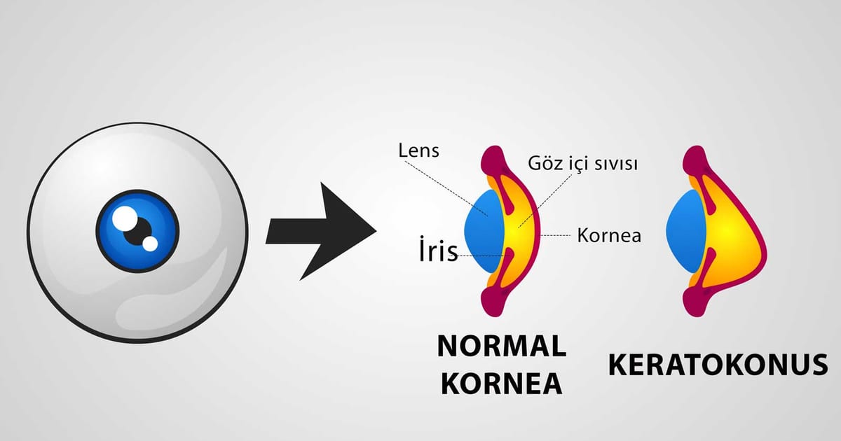
For decades, doctors could correct the blurred vision of keratoconus with lenses but could do nothing to stop the cornea from getting weaker. Corneal cross-linking (CXL) changed that. It is a minimally invasive treatment that strengthens the cornea itself, halting the progression of keratoconus and other conditions that cause the cornea to thin and bulge. Its purpose is not usually to sharpen vision, but to preserve the cornea you have and prevent the disease from advancing toward a transplant.
The science, simply
A healthy cornea holds its dome shape because of strong chemical bonds — "cross-links" — between its collagen fibres. In keratoconus, these bonds are too few and too weak, so the cornea gradually gives way. Cross-linking adds new bonds. The cornea is soaked with riboflavin (vitamin B2) drops and then exposed to a controlled dose of ultraviolet-A light. The light activates the riboflavin, triggering a reaction that forms many new cross-links between the collagen fibres — much like adding rebar to concrete. The result is a stiffer, more stable cornea.
Who is it for?
Cross-linking is recommended for people with progressing keratoconus — most often teenagers and young adults, in whom the disease tends to advance fastest. It is also used for a related condition called post-LASIK ectasia and for some cases of corneal infection or swelling. The key word is progression: the treatment is designed to stop the cornea from getting worse, so it is most valuable when started early, before significant vision has been lost. A stable, long-standing cornea in an older adult may not need it. Suitability depends on corneal thickness and the absence of significant scarring, confirmed by corneal mapping.
The procedure
Cross-linking is an outpatient treatment performed under numbing drops; you are awake and comfortable throughout, and it usually takes under an hour per eye. In the standard "epithelium-off" technique, the thin surface layer of the cornea is gently removed so the riboflavin can soak in, the vitamin drops are applied over several minutes, and the cornea is then treated with UV-A light. A soft bandage contact lens is placed at the end to protect the surface while it heals.
Recovery
The first few days are the most demanding part. As the surface layer regrows, the eye can feel gritty, watery and sensitive to light, and vision is blurry; the bandage lens and prescribed drops make this more comfortable. The surface typically heals within about a week, when the bandage lens is removed. Vision then improves gradually over weeks to months, and it is normal for it to fluctuate during that time. It is important to know that cross-linking aims to stabilise the cornea rather than to improve vision — many people continue to wear glasses or specialty contact lenses afterwards, but with the reassurance that the disease has been halted.
Why it matters
Before cross-linking, a young person with progressing keratoconus often faced a slow slide toward a corneal transplant. Today, a single treatment can frequently stop that progression in its tracks, preserving the cornea and avoiding major surgery. That is why prompt diagnosis and treatment of progressing keratoconus is so important — cross-linking works best when the cornea still has enough thickness and clarity to protect.
This article is general information and not a substitute for a personal examination. If you have keratoconus, please discuss whether cross-linking is right for you with an ophthalmologist.